David B. Durham, M.D.
EXAMINING THE BRAIN’S ‘EXECUTIVE FUNCTION’ WITH Quantitative EEG
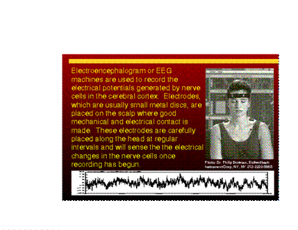
An EEG (Electroencephalograph) is a neurophysiologic technique most often used in the evaluation of epilepsy (or possible epilepsy), but it is also often recommended for headaches, behavioral disturbances, attention disorders, learning problems, language delay, developmental delay, and fainting spells. An EEG is an important test used worldwide to detect neurological abnormalities in children.
The EEG records the ever-present, ongoing electrical activity generated by the neurons in the brain, hence it is also referred to as a "brain wave" test. Abnormal EEG signals include little electrical "explosions" such as the spikes, spike and wave, and sharp waves that are common in epilepsy, however, there are other patterns that indicate disturbances in the brain’s performance.
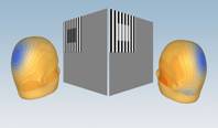
The human brain is so much like a computer circuit board, the nomenclature used to describe many of the parts in computer motherboards are derived from neuroscience. Chemical interactions in the synapse – the “main stage” between neurons (brain cells) form, propagate, and alter the signal received by a particular neuron. It is the neurochemical ‘dance’ in the synapse of neurons that is responsible for how your brain performs: be it mood, concentration, memory or more complicated multisensory tasks involving second to second monitoring and tuning of both motor and sensory information (a good example is flying a helicopter). It is in the synapse, outside and between neurons, where psychotropic medications (“brain active” medications) have their final debut after turning on or off specific genetic sequences inside the neuron itself. The chemical interactions in the synapse are referred to, logically, as “intrasynaptic neurochemical interactions.” All chemical reactions/interactions release energy, partly in the form of electric bursts. Seizures are a classic example of intrasynaptic neurochemical interactions that have gone haywire from either a genetic deficiency, damage from toxins, a chemical problem, or injury to the neurocircuit tissue governing a particular area. We can find the source, or “nidus”, of the seizure in the brain (localizing it to a specific area in a particular lobe of the brain) by measuring the electrical bursts that are occurring throughout the brain created from the on-going intrasynaptic neurochemical interactions in the tissue below. It should be pointed out that there are chemical interactions that produce electricity outside the synapse (again, logically called extrasynaptic neurochemical interactions), chemical interactions in the neurons themselves (logically called intracellular neurochemical interactions), as well as chemical interactions outside the cells (yes, called extracellular neurochemical interactions). It is most convenient for us that the Quantitative or ‘Q’EEG is most sensitive and specific to the intrasynaptic neurochemical interactions, allowing us to specifically target the “main stage” itself – not only where the effect of medications are best measured, but also probably the best place to analyze the chemical phenotype (or chemical result coded by an individual’s unique genetic makeup, or genotype) of a particular region of the brain.
By recording the electrochemical signature of the brain while it is performing “real time” we can characterize and grade it’s perceptual and cognitive operations, and form an accurate model of how particular areas are performing. Our approach places emphasis on multisensory processing, selective attention, task-switching in divided attention, error correction and “pre-planning” functions of the frontal lobe of the brain. These operations were selected because they require the cooperation and engagement of all the functions required of the frontal lobe. It is the area that is most involved in coordinating and planning, working memory, concentration, focused and divided attention, navigating ambiguous or novel tasks, and impulse control. By measuring how the brain is performing in these areas by recording ‘real time’ QEEGs, we achieve a degree of diagnostic sensitivity that far exceeds any other procedural or imaging modality. This markedly enhances our diagnostic certainty in neuropsychiatric disorders—from Attention Deficit Hyperactivity Disorder (ADHD) to atypical cognitive problems seen in traumatic brain injury, to cognitive problems specific to major depression and those not only in ‘dementia’ but specific to the different subtypes of dementia (e.g. Alzheimer’s Disease vs. Frontotemporal Dementia vs. Parkinson’s Dementia) as well as cognitive impairment seen in Multiple Sclerosis, Wilson’s Disease, and stroke. In short, the gold standard for the temporal — or “real-time” — performance of the brain is the QEEG.
Dr. David Durham is Director of Neuropsychiatry for the Mosaic Neuroscience Group in Santa Fe, New Mexico, and a Clinical Assistant Professor of Psychiatry at the University of New Mexico, School of Medicine.
